TETRODES FOR MONKEYS
J. S. Pezaris, M. Sahani, R. A. Andersen
Division of Biology
Computation and Neural Systems
California Institute of Technology, Pasadena, CA 91125, U.S.A.
August, 1995
NOTE: This paper is a slightly modified version of a paper by the same title and authors which will appear in the proceedings of CNS*95.
ABSTRACT
We have adapted the McNaughton-Wilson tetrode for use in monkey cortex. Eleven penetrations have been made over macaque area MT to evaluate the mechanical and electrical properties of our design. Experiments have been made with 15 and 25 micron insulated nichrome wire running through oil-filled 33 gauge stainless steel guide tubes. Electrically, the tetrodes compare favorably with traditional tungsten electrodes for single-unit isolation, and are superior for multi-unit recordings. Preliminary histology shows straight tracks at up to 12 mm tetrode extension.
INTRODUCTION
The primary contribution of this paper is to show that the McNaughton-Wilson tetrodes [1] can be used to successfully record from monkey cortex, and to detail the parameters and techniques used in that process. The design of our tetrodes is presented, including specific construction details, followed by the description of experiments performed to measure their characteristics. We close with a list of intended design modifications and follow-up experiments.Tetrodes and Spheres of Sensitivity
A tetrode is a bundle of four individually insulated fine wire electrodes, whose tips lie closer together than their respective spheres of sensitivity. An exact analogy can be drawn between neural recordings obtained from such a device and quadraphonic musical recordings. A neuron that lies in the overlap of two or more of these spheres is detected by two or more electrodes. Assymetries, whether in electrode construction or relative geometry, insure that different electrodes record this neural signal through slightly different filters. The comparison of signal across channels allows the disambiguation of signals that appear identical to a single electrode. Later in the paper, a tetrode recording is presented which shows this effect.
Animal Preparation
Previous reports describing tetrodes have concerned themselves with rat [1] or cat [2] preparations. Two features of our awake monkey preparation constrained our design. First, while a recording chamber is chronically implanted, the surface of the brain is exposed and electrodes are inserted and removed during each recording session. Thus, the dura mater must remain intact and is subject to toughening. Second, the brain of the macaque is larger than those of either the rat or the cat and profoundly gyrated in comparison. Thus even cortical areas can lie quite far from the exposed surface. A mechanism is therefore required to penetrate the dura and deliver the delicate tetrode wires to relatively deep neural structures, while minimizing tissue insult. Our approach is described below.
We initially chose a method which uses a carrier tube to hold the tetrode wire within the brain (Mk I), combined with a normal guide tube to puncture the dura. A modified design under development combines the two functions (Mk II). These two designs are described in detail below.
METHODS
Tetrode Design, Mk IThe basic design is a four-conductor twisted electrode running in a carrier tube. 15 or 25 um insulated nichrome (Reid or Sigmund Cohn, respectively) is used to construct the tetrode bundle. To penetrate dura, a 21 gauge stainless-steel guide tube is used, inside of which a carrier tube assembly tapers to a 33 gauge oil-filled cannula holding the tetrode bundle. Conductive silver paint is used to electrically and mechanically connect the wires to a four-pin connector mounted on the carrier tube. Thus, the tetrode bundle runs from the recording end, up through the carrier tube, and exits in a loop which terminates on the connector. See Figure 1. Construction is described in the following paragraphs.
The tetrode wires are twisted in a motorized jig. Once twisted, the four wires are glued together using cyanoacrylate glue, allowed to dry, carefully cut free of the jig, and set aside during the construction of the carrier tube.
The carrier tube is constructed by gluing three lengths of cannula, 9 cm of sharpened 33 gauge, 8 cm of sharpened 26 gauge, and 1.5 cm of 21 gauge, flush at the blunt ends with quick-setting epoxy. The four-pin connector is glued transversely to the outer cannula. The assembly is designed to withstand gentle handling and clamping in the microdrive while providing a small tip cross-section to minimize tissue insult. See Figure 1.
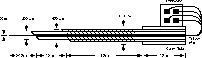
Figure 1: Tetrode Mk I, section
This carrier design is extended a few millimeters beyond the end of a guide tube (not shown) which has punctured the dura. The tetrode bundle, free to move inside the oil-filled center channel, is advanced into the area under investigation. Drawing not to scale. Click here or on the drawing for an enlarged view.
Once the glue has set, the carrier tube is reamed with music wire, cleaned with acetone, and filled with silicone oil. Oil-filling the carrier tube is important to increase construction yield and reduce the chances of kinking during use. The tetrode bundle is loaded into the carrier tube, and the free ends carefully attached to the four-pin connector using conductive silver paint (Silver Print, GP Electronics, Rockford, IL). No step is taken to remove the wire insulation: it is thought to dissolve in the conductive paint vehicle. The recording end of the tetrode bundle is cut blunt with fine scissors and plated in 5% gold chloride (AuCl4) solution for approximately 35 uA-s per tip. This yields a 1 kHz tip impedance of 0.2-1.5 Mohm at plating time, as measured on a BAK impedance tester.
Tetrode Design, Mk II
Our improved design, incompletely tested in the current experiment, simplifies use and increases reliability. Three nested lengths of cannula are again glued to form a carrier tube. The tetrode bundle is attached to a second sleeve of layered cannula which forms a cap that slides over the lower carrier. With the tetrode withdrawn, the carrier is advanced through the dura but short of the brain, and the tetrode extended under hydraulic control. This approach obviates the need for a separate guide tube, as required for Mk I. See Figure 2.
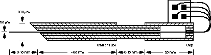
Figure 2: Tetrode Mk II, section
The carrier tube punctures the dura directly, but is not advanced into the brain. The cap is advanced over the carrier, forcing the tetrode wire, which has been fixed to it, into the brain. Drawing not to scale. Click here or on the drawing for an enlarged view.
Equipment
Our experimental setup is as follows. A hydraulic microdrive (Fred Haer Corp, Brunswick, Maine) is used to position the tetrodes. Tetrode signals are amplified by a custom four-channel headstage amplifier (A=100) feeding a custom four-channel variable-gain preamplifier (A=1 to 5000, nominally set to 500). The preamplifier feeds anti-alias filters (f_c=6.4 kHz, Tucker-Davis Technologies, Gainsville, Florida) and four-channel instrumentation-grade A/D (f_s=51.2 kHz, decimated to 12.8 kHz, also TDT). Data are collected on an IBM-PC compatible and analyzed on Sun computers using custom software.
Experiment
In this experiment, we sought sought to verify that recordings could be made using this technology in semi-chronic monkey preparations. Subsequent to this verification, the following issues were explored: wire size, tip plating, penetration straightness, and tissue insult.
Eleven penetrations were made over the period of a week. Six penetrations were made with 15 um wire tetrodes, three with 25 um wire tetrodes, and two with traditional tungsten electrodes as controls. All penetrations were made in the left hemisphere of monkey 92-13 in an awake preparation. Penetrations were made to tetrode extensions of 4-12 mm, limited by variations in the mechanical designs of the four tetrodes used. Although recordings were made from all penetrations, electrical problems confounded the recordings from two. Some penetrations were made without leaving lesions, others had a pair of electrolytic lesions made 2 mm apart, and others had lesions made every 500 um; all lesions were made during electrode withdrawal, after recording had finished. After the last day of recording, the brain was perfused using standard techniques.
RESULTS
An example tetrode recording is shown in figure 3. This is of particular interest because it clearly shows one unit (a) detected on channel 3, a second unit (b) on channel 2, and both on channel 1 at approximately equal amplitude, illustrating the monaural-versus-quadraphonic analogy suggested earlier. Two additional units (c, d) were detected in this recording. Notice how similar three of the four waveforms are on channel 1, making it very difficult to disambiguate between them given only the information from that trace.
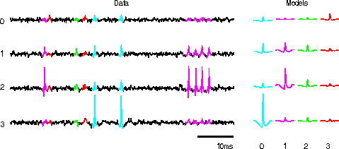
Figure 3: Example Tetrode Recording
On the left is a short stretch of the four channels of data. Heavier sections represent detected events. Letters signify events from four presumably different units. On the right are the averages of all events from each of the four identified cells.
Electrical Performance
Analysis of tetrode electrical performance was primarily comparative. Signal-to-noise ratios of approximately 14:1 were found for single-unit isolation. In situ impedance at 1 kHz was consistently 0.5-0.7 M ohm for plated electrodes, 1.0-2.1 M ohm for non-plated electrodes. This compares to 23:1 and 0.8 M ohm for the two control penetrations. Non-rigorous examination of recorded data reveals multiple distinct cells per stable recoding location for tetrodes.
Wire Diameter
No significant differences have been found between the various recordings or penetrations based on wire diameter. Further exploration may be necessary to clearly elucidate any distinctions.
Tip Plating
Plating the electrode tips is essential to producing good recordings. This is clearly depicted by comparing the spectral impedance of an unplated and plated tetrode tip, as seen in Figure 4. This is an area of active pursuit.
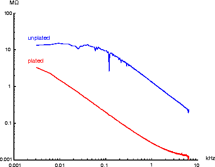
Figure 4: Impedance Curves
This graph shows a spectral analysis of tip impedance for a typical tetrode wire. The upper curve is from a freshly cut tip in saline (the spike at 120 Hz is due to power line interference). The lower curve is from the same tip after gold plating.
Penetration Straightness and Tissue insult
We have histologically identified all marked penetrations and have determined that the tetrode wires ran straight and the tracks are not unusual. The deepest tetrode penetration, some 12 mm, included 12 lesions which lie in a straight line, even at the maximum extension. Preliminary comparison of tracks between the control penetrations and the two wire diameters show that all three are comparable. That is, the tetrode wire caused no additional tissue insult.
SUMMARY
We have described the design and construction of a tetrode design appropriate for use in the chronic monkey preparation. Experiments were performed to verify the mechanical and electrical performance of the tetrodes which showed them to be functional and adequate for simultaneously detecting multiple neighboring cells in monkey cortex.
REFERENCES
[1] Wilson, M. A. and McNaughton, B. L., ``Dynamics of the Hippocampal Ensemble Code for Space,'' Science, 261(5124) pp. 1055-1058 (1993).[2] Gray, C. M., et al, ``Tetrodes markedly improve the yield and reliability of multiple single unit isolation from multiunit recordings in cat striate cortex,'' Society for Neuroscience Abstracts, 20(1) p. 625 (1994).
John Pezaris
Caltech
Mail Code 216-76
Pasadena, CA 91125
pz@caltech.edu 17 September 1995